The cell wall provides a rigid form to the cell.
It is unique to plants.
Animals do not have a cell wall.
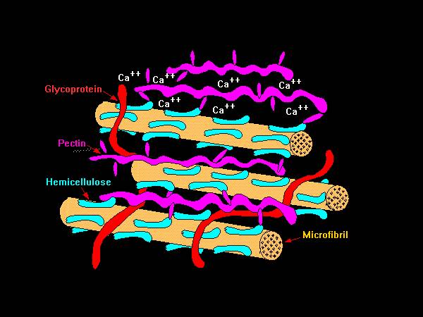
The primary cell wall is made up of :
- Cellulose
- Hemicellulose
- Pectin
- Glycoproteins
A schematic representation of the components of the primary cell wall.
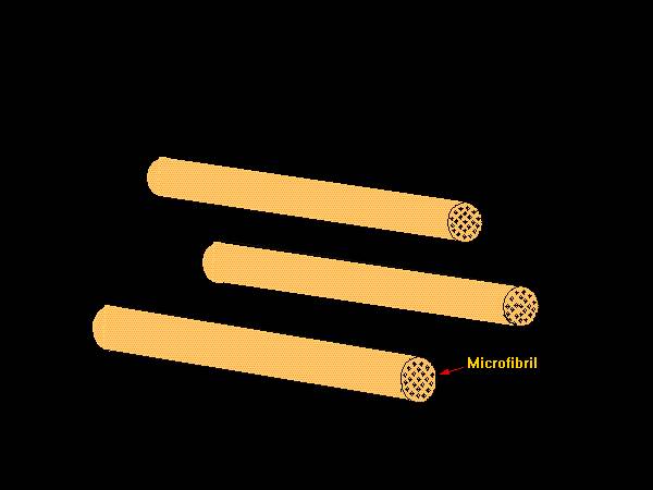
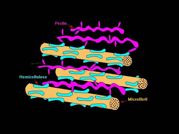
The main component of the cell wall are the cellulose microfibrils.
Microfibrils are groups of cellulose molecules held together in long chains.
A schematic representation of the components of the primary cell wall.
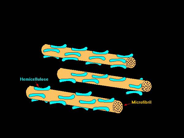
Hemicellulose is the next most abundant material in the cell wall.
Hemicellulose molecules are bond to the surface of the microfibril.
A schematic representation of the components of the primary cell wall.
Hemicellulose molecules are cross-linked to pectin molecules.
Pectin acts like a glue to hold the molecules together.
A schematic representation of the components of the primary cell wall.
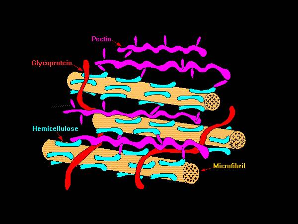
Glycoproteins and certain enzymes are also part of the cell wall.
They are between microfibrils.
A schematic representation of the components of the primary cell wall.
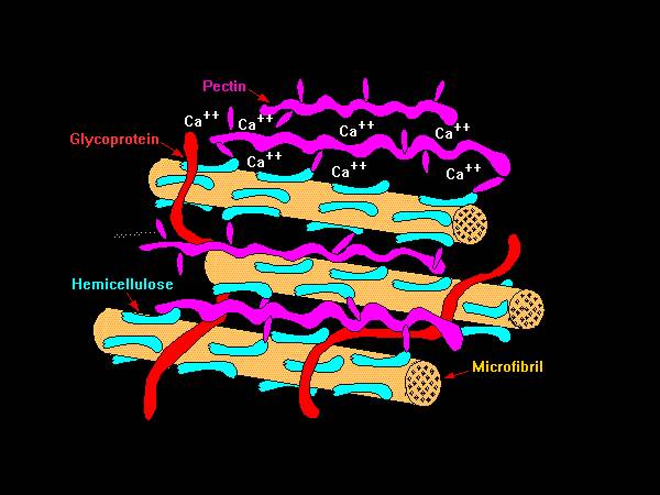
Calcium in the cell wall act as bridges between pectin molecules.
They add strength to the wall.
A schematic representation of the components of the primary cell wall.
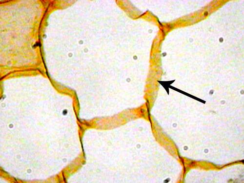
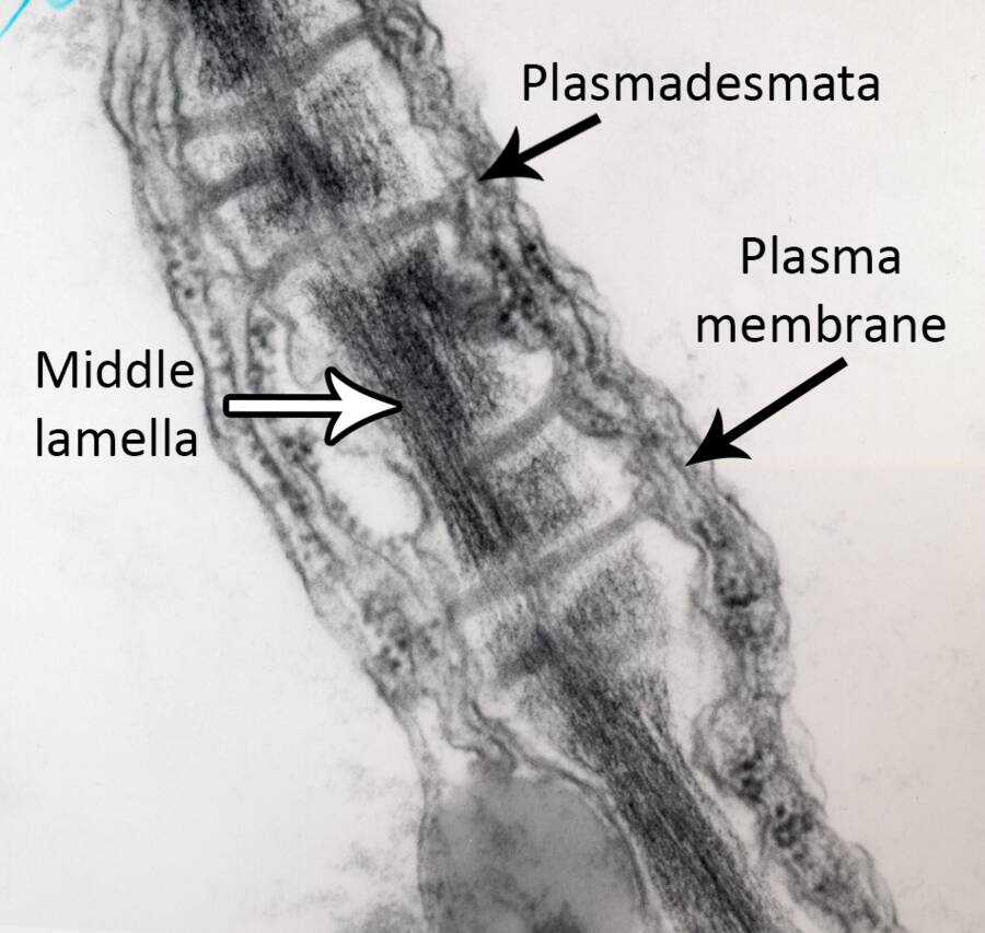
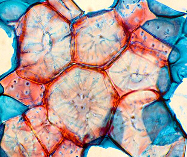
Cells are interconnected by pores in the cell wall through which the plasma membrane extends.
These are the plasmadesmata.
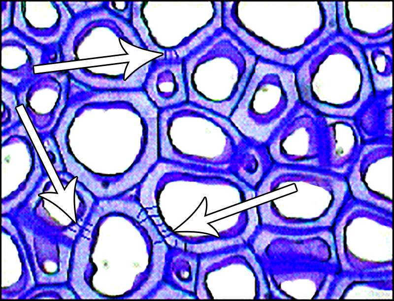
Here is an electron micrograph of primary cell walls between two cells.
The arrows show plasmadesmata extending through the cell walls and the electron dense middle lamella.
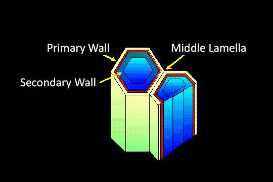
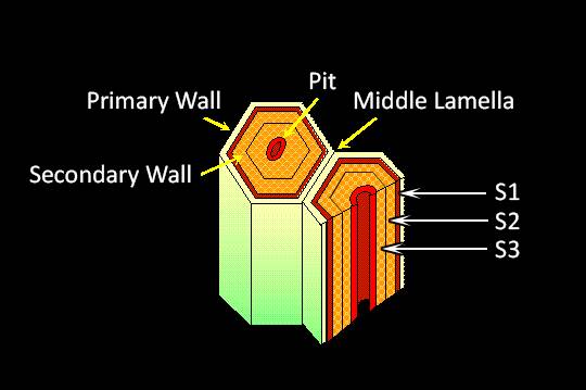
Some cells mature to have secondary cell wall thickening.
The cell wall grows to the inside of the cell.

The primary cell wall is relatively thin and flexible and can expand.
It has a large vacuole.

The initial secondary cell wall forms under the primary wall.
With the deposition of lignin, the secondary wall becomes thick and rigid.
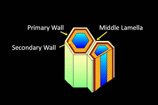
Eventually, the cell loses its cellular components and a pit forms within the center of the cell.
These cells with full secondary cell walls are dead at maturity.
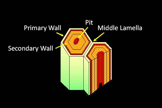
The secondary wall can have up to three layers called S1, S2, S3.
Types of cells with secondary cell walls include xylem, sclerieds and fibers.
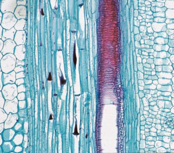
Xylem
Sclerieds
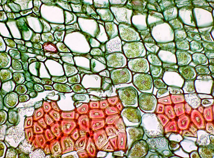
Fibers