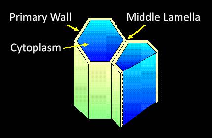
Collenchyma cells have primary walls that are usually thickened especially at corners or edges.
They function for structural support.
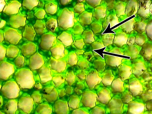
Collenchyma cells are characterized by having a thickened primary cell wall that is not lignified. The thickening takes place in the middle lamella.
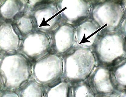
It is easy to see the cell wall thickening between each collenchyma cell in celery.
The middle lamella is evident as an "X-shaped" mark between cells.
Cross-section of a celery stem.
Compare the appearance of the thickened primary walls of collenchyma cells with the thickened secondary walls a fiber cell that contain lignin.
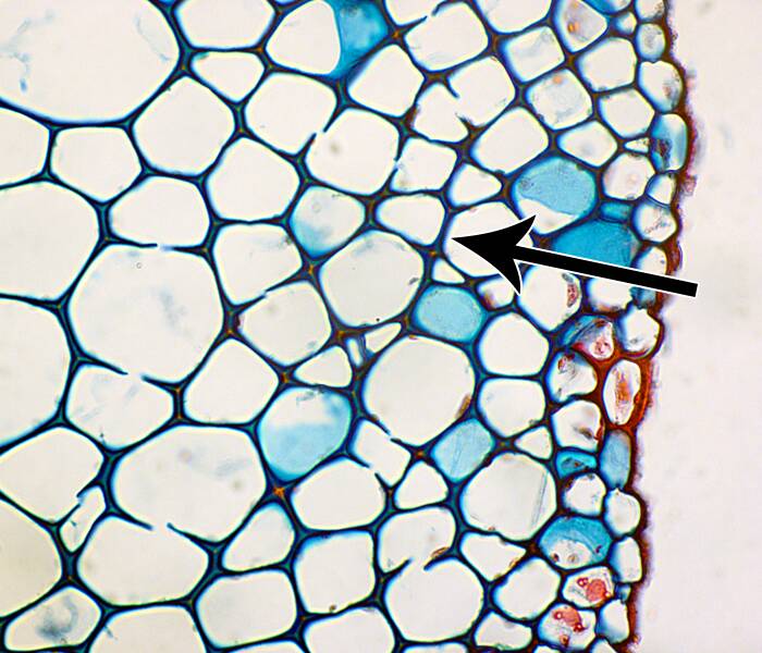
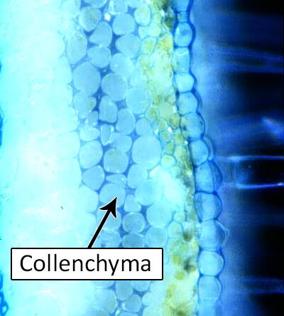
Collenchyma cells with the cell wall stained dark blue.
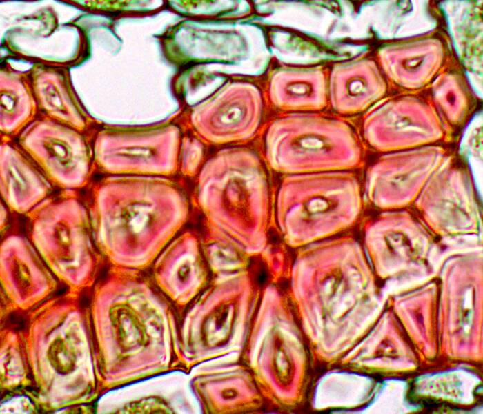
Fiber cells with the entire cell wall stained red.
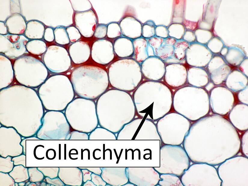
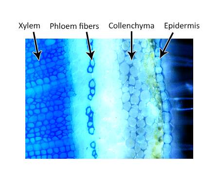
Collenchyma cells are also usually found in groups under the epidermis.
Cross-section of a tomato stem.
Collenchyma cells and fibers both function to support the stem or leaf, but unlike fibers, collenchyma cells are usually living at maturity.
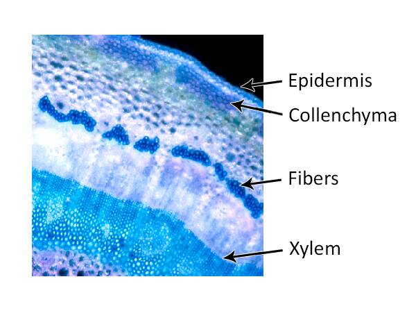
Cross-section of English ivy (Hedera helix).