Parenchyma are diverse cells and can have many different shapes and be very specialized in their function.
Parenchyma cells are usually isodiametric or polyhedral in shape.
They have only a primary cell wall and retain the ability for future cell division.
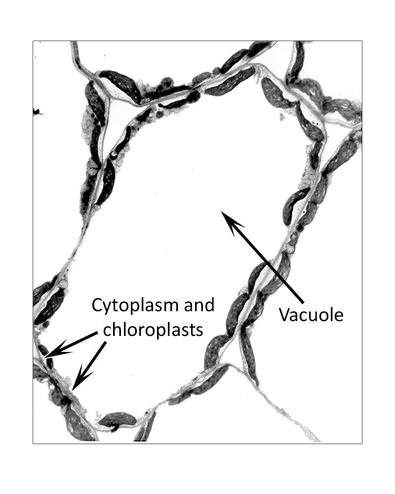
Parenchyma cells contain a nucleus and when they are first formed, they are densely cytoplasmic and have several small vacuoles.
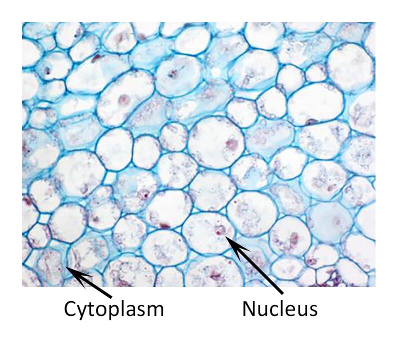
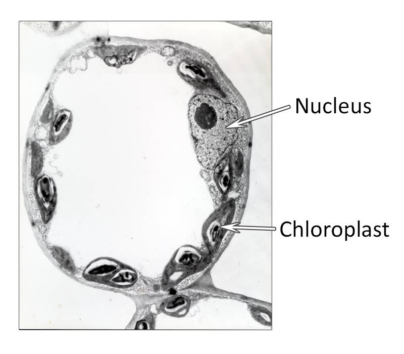
However, in older parenchyma cells the vacuoles merge into one large central vacuole with the cytoplasm and organelles - like these chloroplasts on the edges of the cell.
Parenchyma are diverse cells and can have many different shapes and be very specialized in their function.
Based on their function specialized parenchyma cells can grouped as:
- Ground tissue
- Storage tissue
- Chlorenchyma
- Aerenchyma
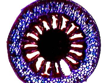
The cortex and pith in stems and roots are parenchyma cells commonly referred to as the ground tissue.
The ground tissue surrounds the vascular system in the stem.
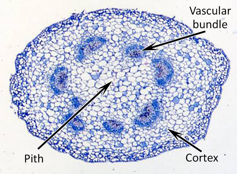
Stem cross-section
There are several distinct storage tissues in plants. Three examples are:
- Seed endosperm
- Storage parenchyma
- Ray parenchyma
Storage parenchyma in cotyledon.
The endosperm is designed to store food for the developing embryo and seedling.
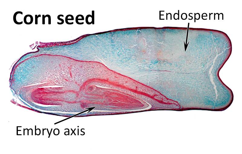
The endosperm is filled with lipid bodies, protein bodies and especially amyloplasts containing starch.
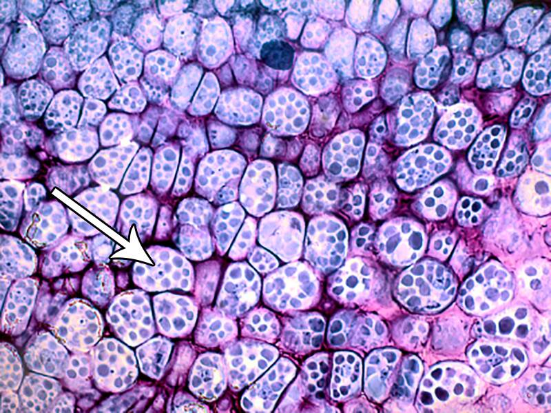
Protein bodies in legume endosperm.
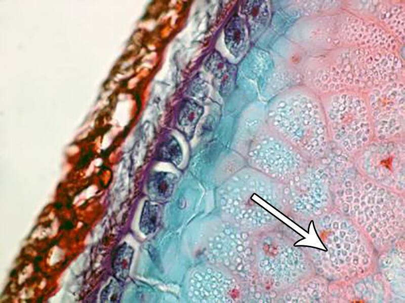
Amyloplasts in grain endosperm.
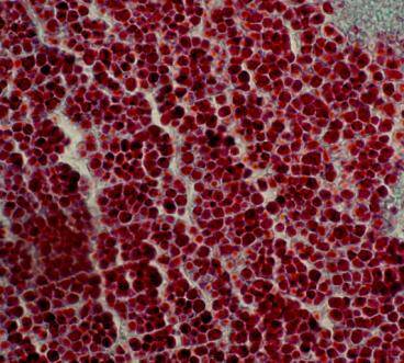
One of the major functions of parenchyma tissue is to store food reserves.
In some cases, large amounts of parenchyma are used for storage as in this beet root.
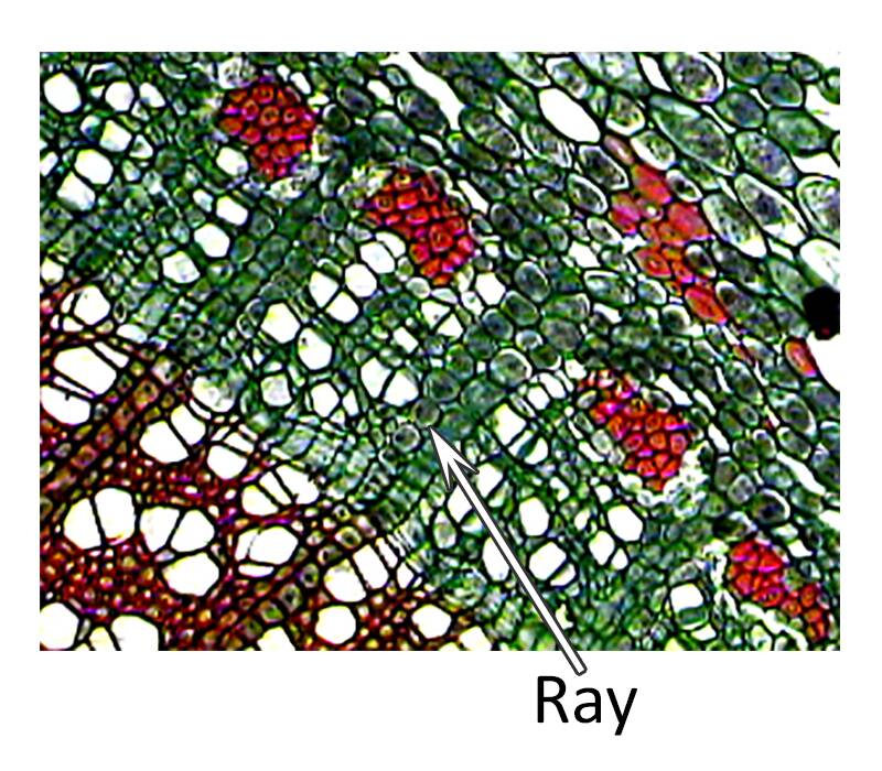
A second major parenchyma type used for storage is ray parenchyma.
Ray parenchyma cells grow horizontal to the developing stem, sometimes deep within the non-living xylem cells.
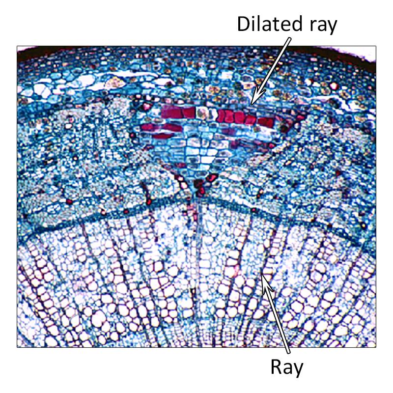
Ray cells are an important storage tissue to store carbohydrates and proteins over the winter in stems.
These stored materials are used to support new spring shoot and leaf growth.
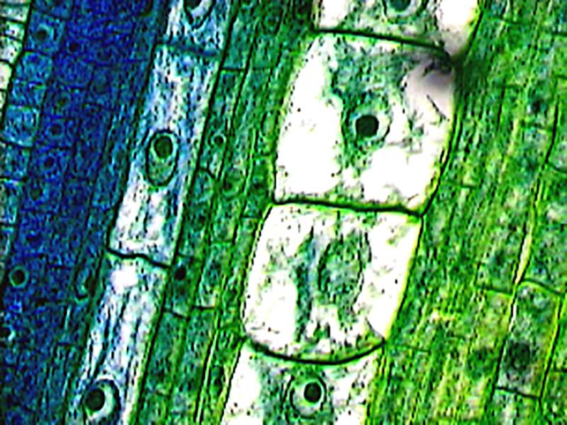
Parenchyma cells in the leaf actively involved with photosynthesis are termed chlorenchyma.
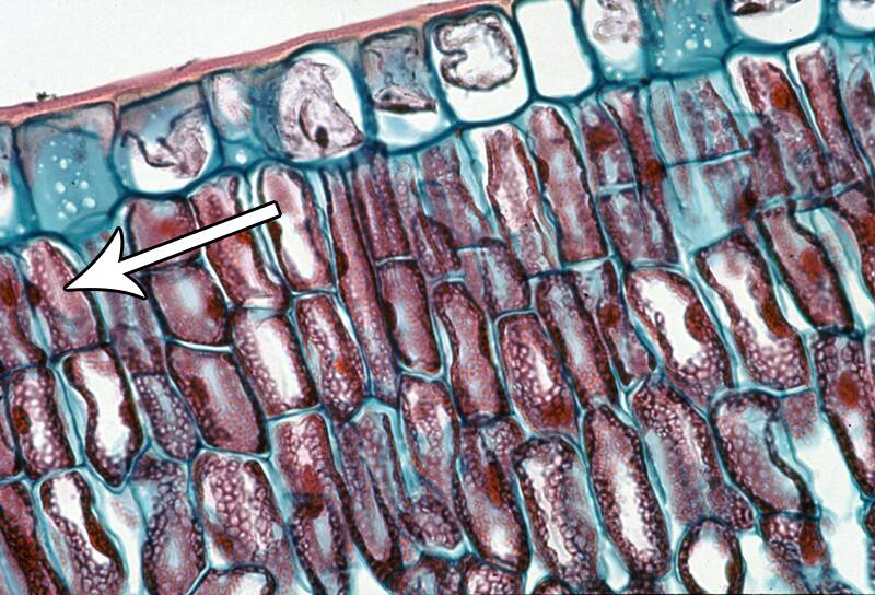
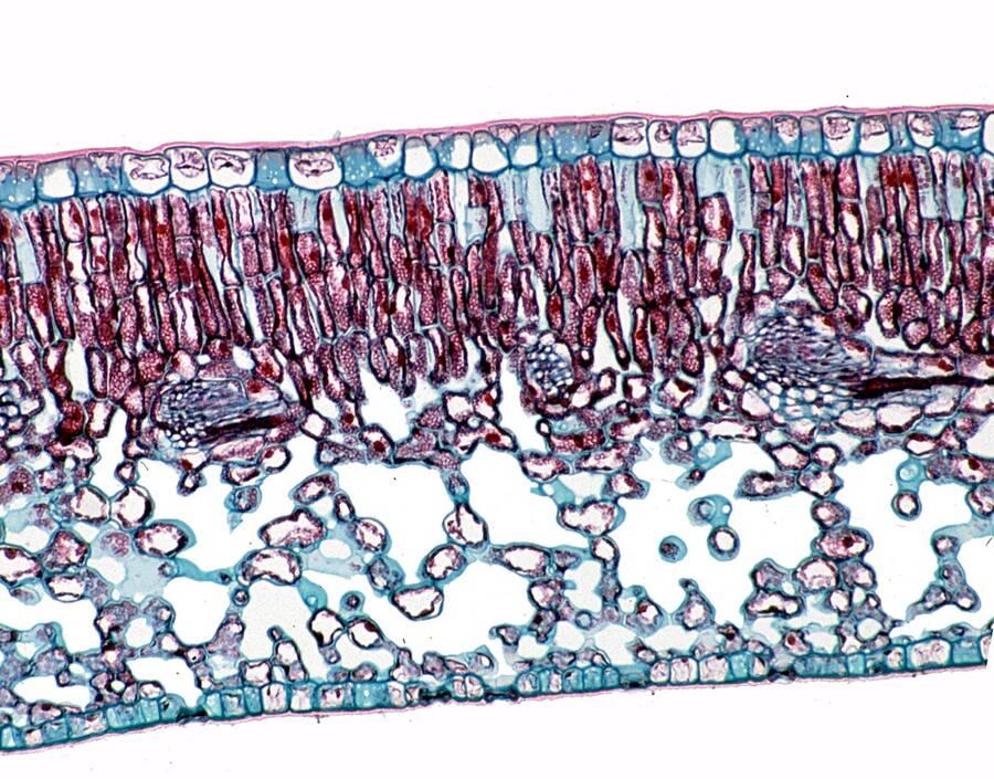
Chlorenchyma can be divided into the leaf palisade layer and the spongy mesophyll cells.
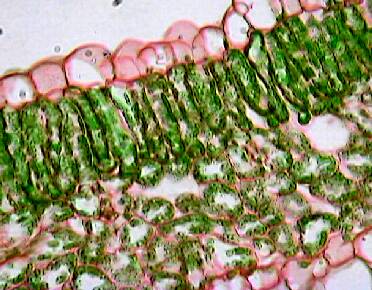
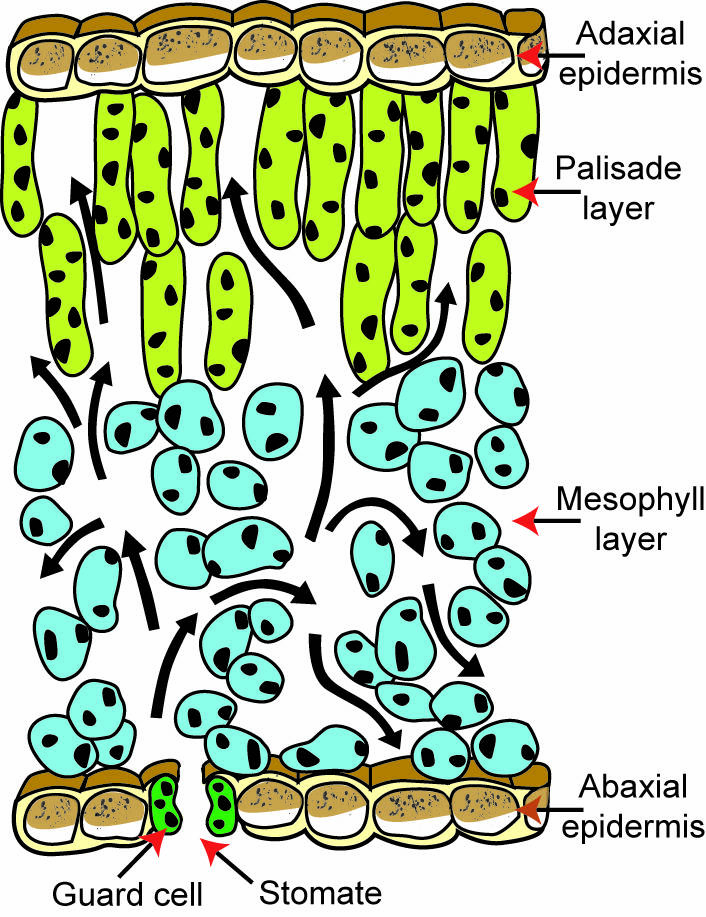
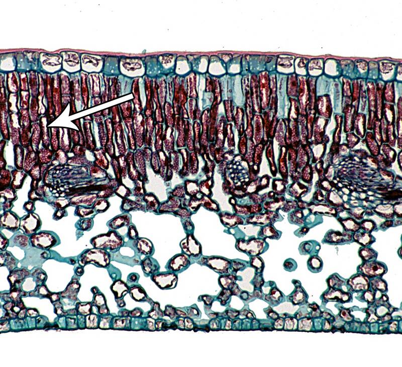
The palisade layer is usually one to three cell layers deep and develops under the epidermis on the top (adaxial) side of the leaf.
The spongy mesophyll cells occur below the palisade layer and are loosely packed together.
This creates air chambers that allows carbon dioxide to move from the stomata on the underside of the leaf to these chloroplast containing cells.
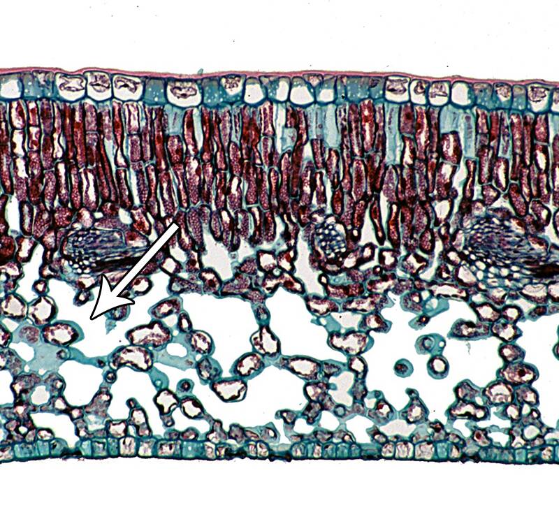
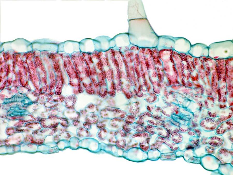
The leaf Palisade and Mesophyll cells contain abundant chloroplasts easily seen from the red color of chlorophyll in the leaf cross-section below.
This electron micrograph shows a single mesophyll cell with an active nucleus and numerous chloroplasts.
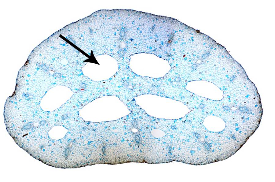
Aerenchyma can occur in leaves, stems or roots and act as air passages.
They occur commonly in water logged roots and in aquatic plants.
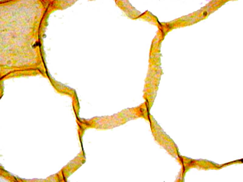
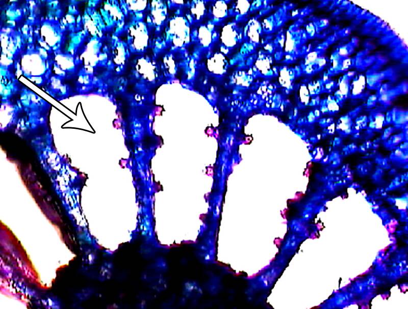
Aerenchyma is easily seen in the leaf of the floating water lily.
Air in arenchyma spaces help the leaf to float.
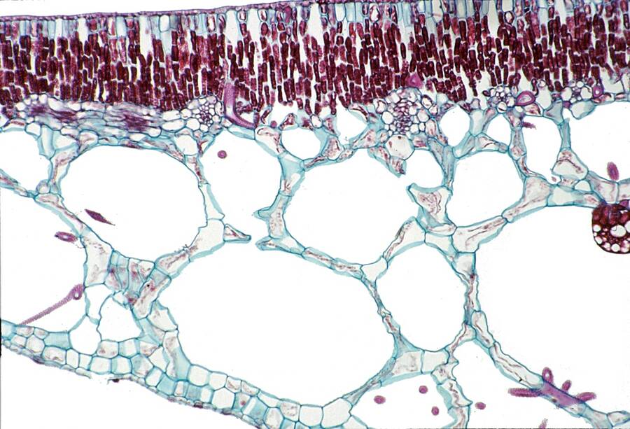
Leaf cross-section in Nymphaea (Water lily)
Aerenchyma can form from elongating cells creating large intercellular spaces or by cells disintegrating to leave a large air space.
Stem cross-section in Myriophyllum (Parrot feather)
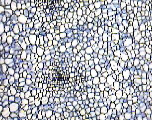
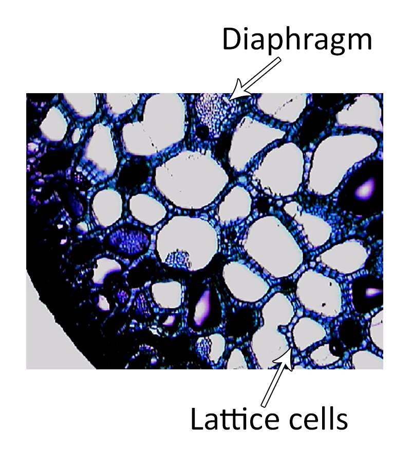
Small cells form an intricate lattice structure creating aerenchyma in water hyacinth.
These air passages form pipes for air transport. However at intervals, cellular diaphragms form that allow air but not water to move through the passages.
This prevents sinking of this floating aquatic plant in case of damage that would allow water into the air passage.
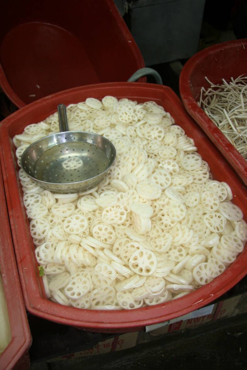
Lotus (Nelumbo) produces an edible root with large arenchyma spaces.
They help move air down into the submerged roots.