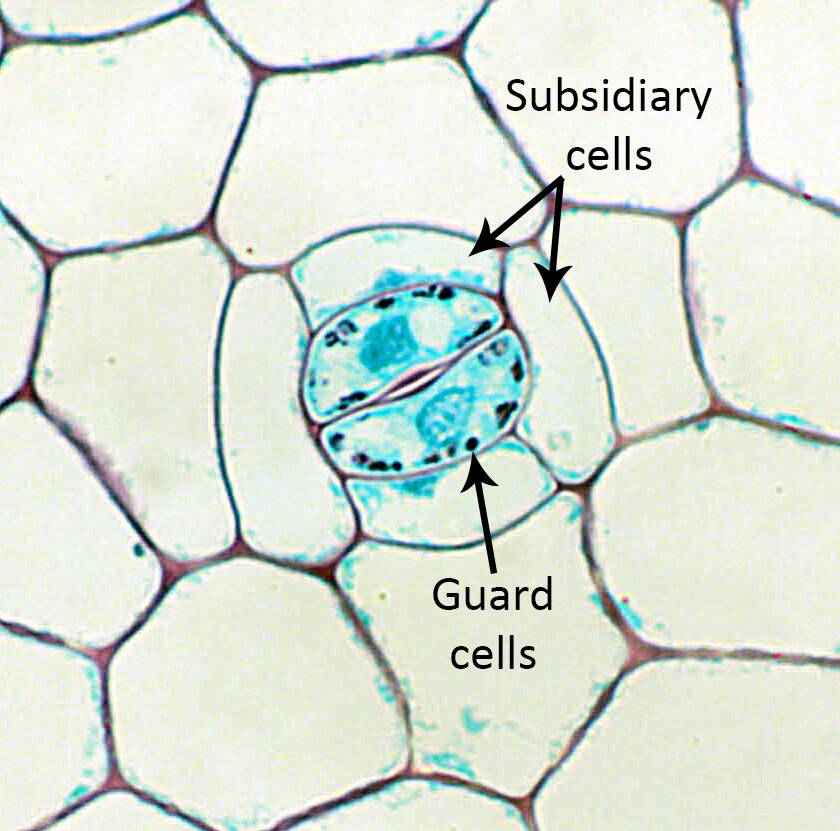
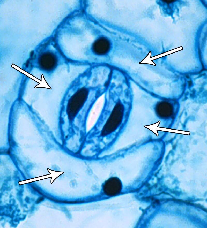
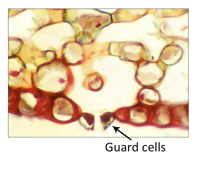
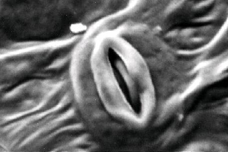
A stomate is a group of cells in the epidermis made up of guard cells and subsidiary cells.
Stomata are designed to allow gas exchange.
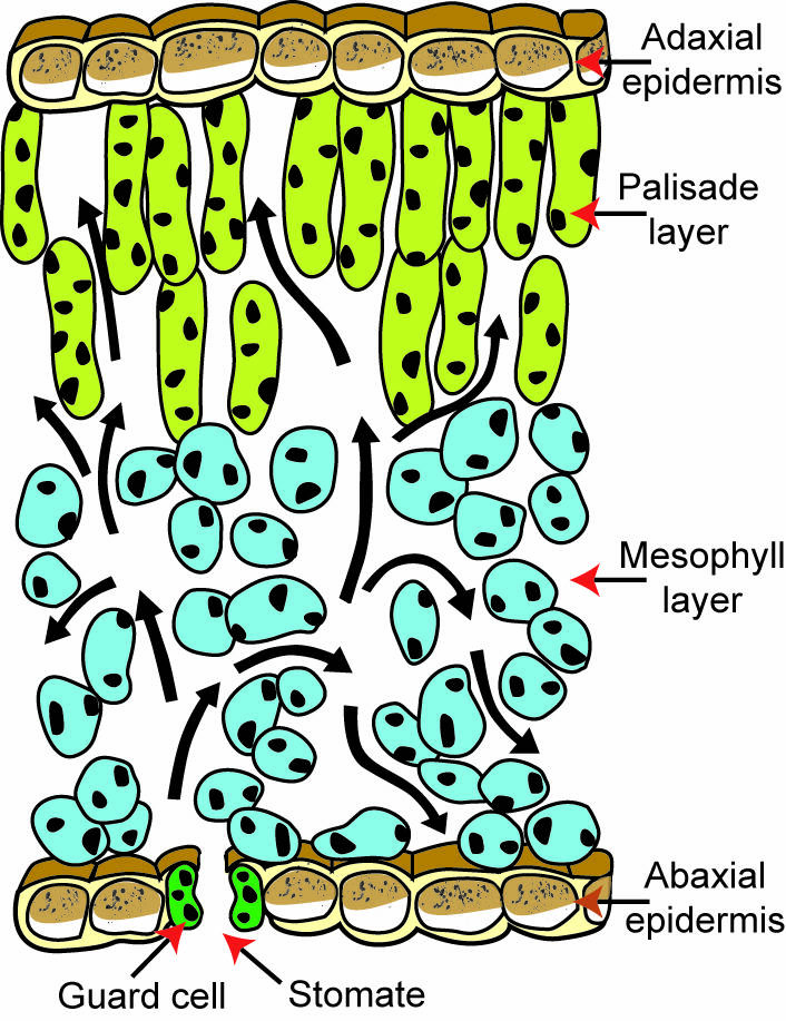
Stomata are most frequently found on the bottom (abaxial) side of leaves, but may occur on the top (adaxial) of leaves, stems and even fruit.
A stomatal complex consists of:
Guard cells
and
Subsidiary cells.
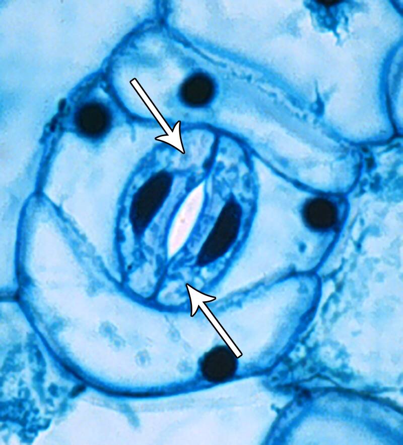
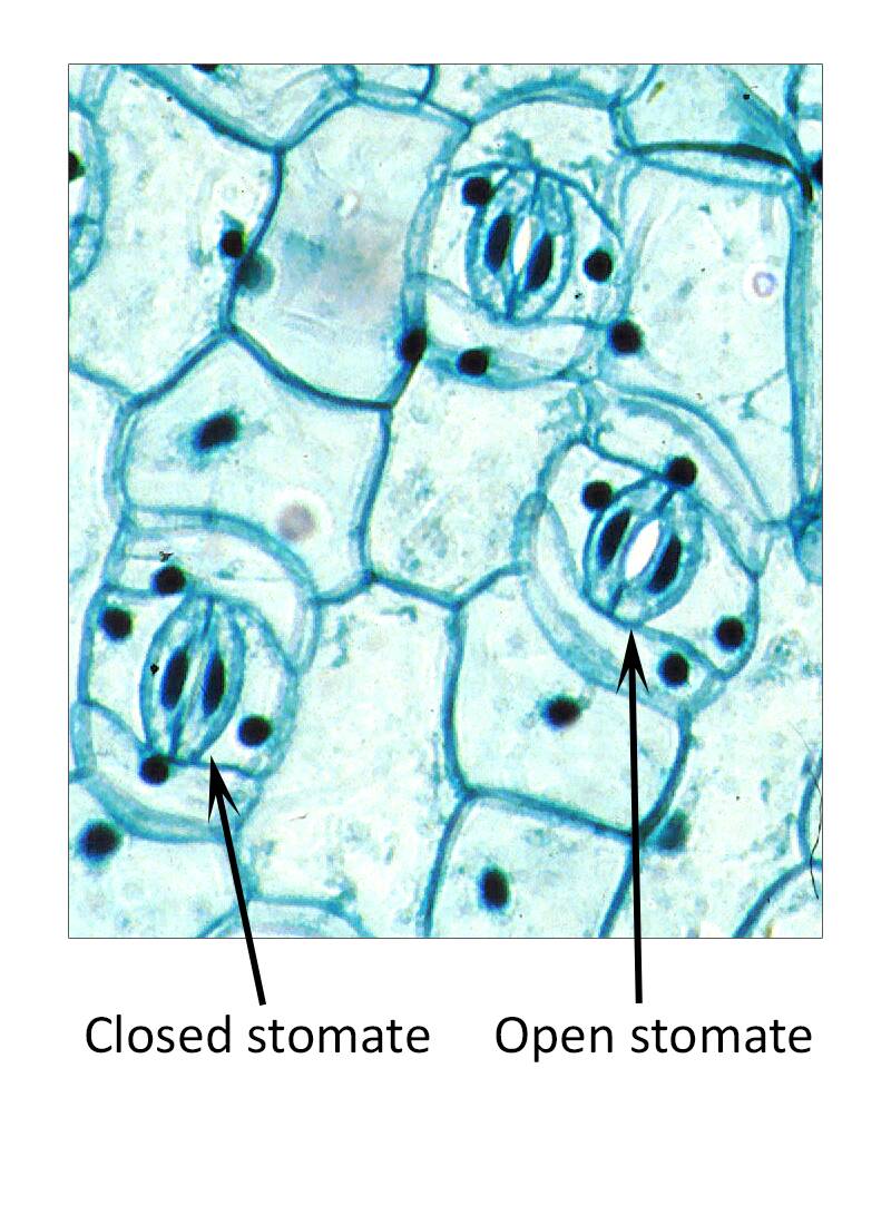
Guard cells are a pair of kidney-shaped cells that form the opening of the stomate. When the guard cells are turgid (full of water), they are open and allow gas to enter the stomate.
However, this also exposes the leaf to potential water loss.
When the guard cells are flaccid (less water), they are closed to prevent air exchange and water loss.
Subsidiary cells - These are cells bordering guard cells of the stomate.
They do not directly participate in the opening and closing of the stomate, but may aid it in their functioning.
These cells vary in their arrangement and pattern depending on the plant species.
When the plant is turgid (full of water) the guard cells are swollen and the stomate is open. This allows carbon dioxide to enter the leaf for photosynthesis, but it also allows water to leave the opening. Water leaving the leaf is called transpiration.
During times of water stress, the guard cells lose water and shrink. This closes the stomate. Most stomata are also closed at night, since most plants do not need to fix carbon dioxide in the dark and the plant does not need to lose water needlessly.
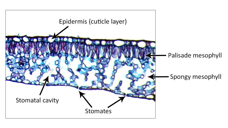
In side view, the stomatal cavity is evident.
The stomatal cavity helps with gas exchange across the leaf.
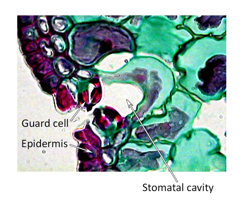
This is a stomatal cavity in pine. This is an example of a sunken stomatal cavity.
Guard cells are not directly on the epidermal surface.
This arrangement is more efficient at preventing water loss.
A photomicrograph and electron micrograph of the guard cells.
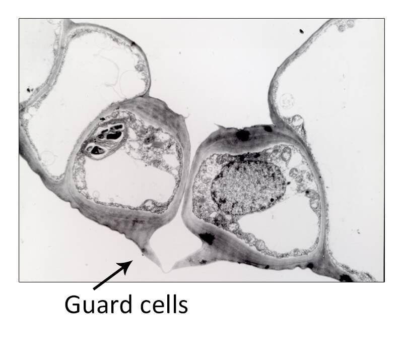
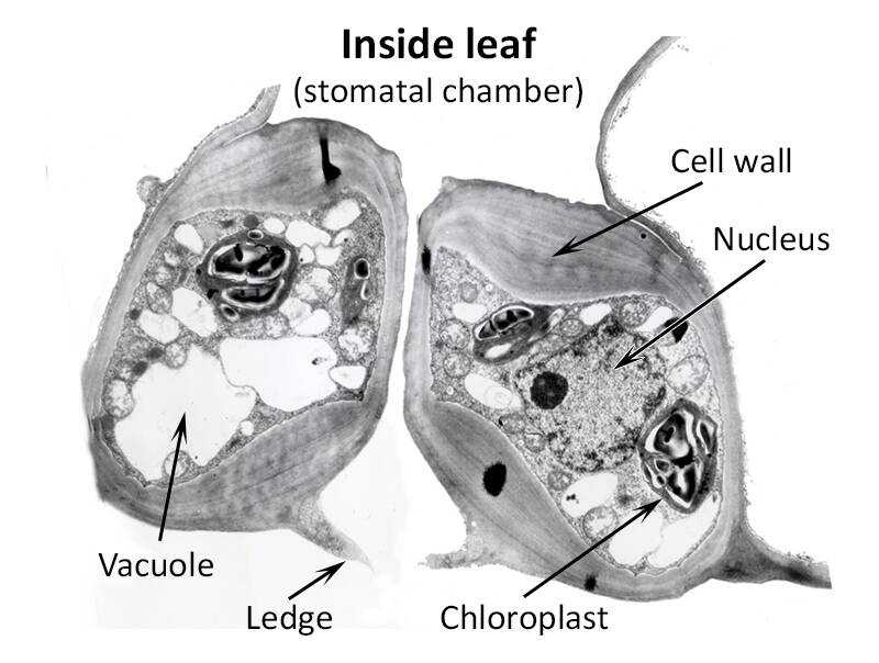
An electron micrograph of the guard cells reveals the large nucleus, relatively thick cell walls, numerous vacuoles and chloroplasts.
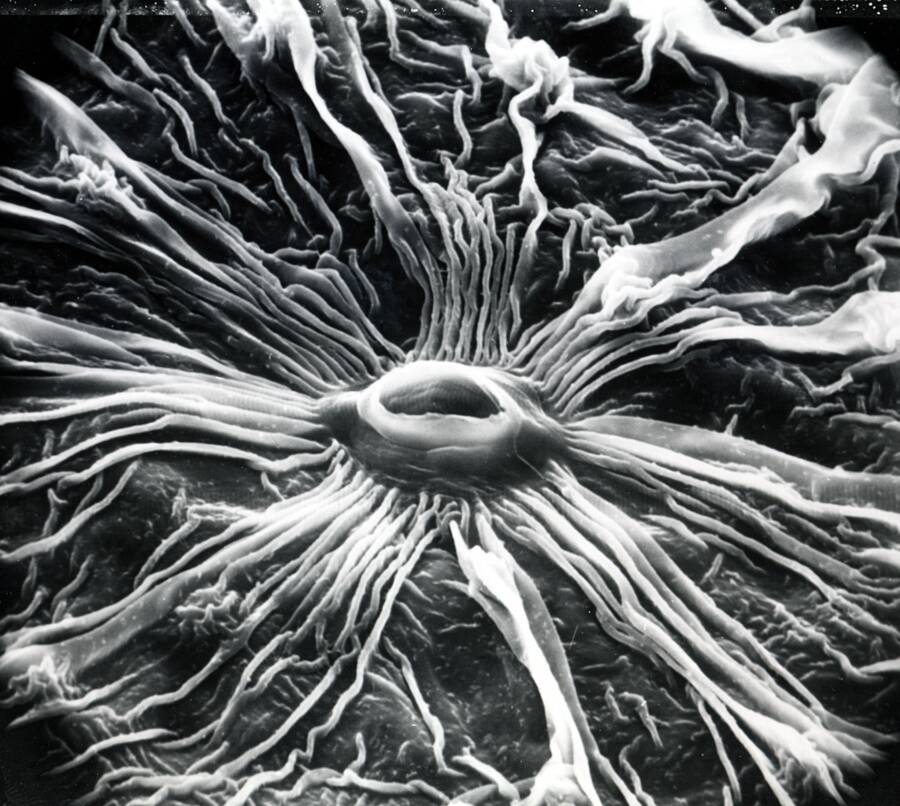
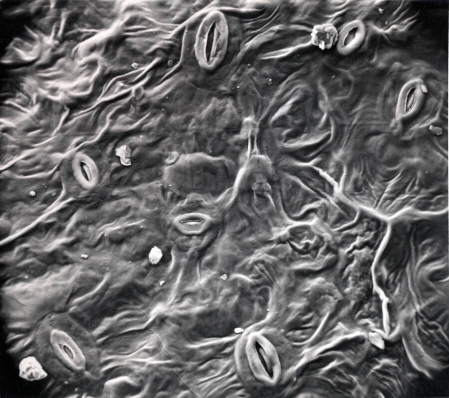
Surface view of a typical stomate by scanning electron microscopy.
Electron micrograph of stomate in dogwood shows a wonderful surface pattern over subsidiary cells made by strands of wax.
Wax on the epidermal cells helps to reduce water evaporation from the surface.