The endoplasmic reticulum or ER is located in the cytoplasm.
It is a membrane system that extends throughout the cell.
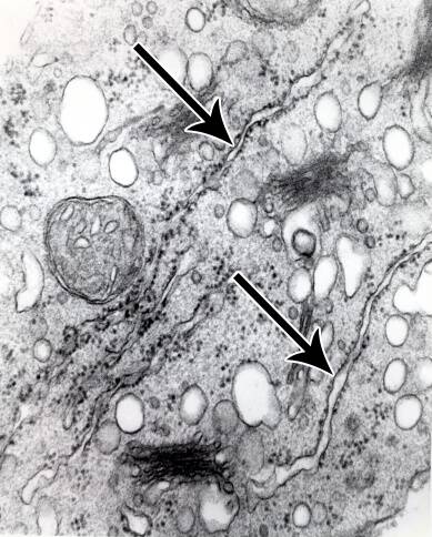
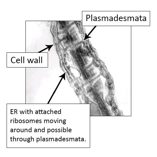
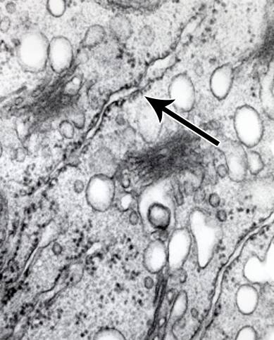
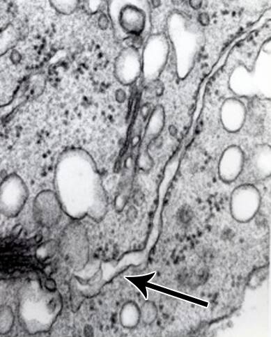
In electron micrographs, the ER appears as two parallel membranes.
The endoplasmic recticulum is the source for the membrane material used to create the vacuole, Golgi bodies and microbodies (like protein bodies and glyoxysomes).
The ER is also thought to function in cellular communication.
ER are found associated with the nuclear envelope and are even seen extending through the plasmadesmata between cells.
There are two types of ER
Rough ER have ribosomes attached to them and are actively involved in protein synthesis.
Smooth ER are not associated with ribosomes.