Golgi body (also called the dictyosome) is a group of stacked membranes responsible for depositing material to the cell wall.
Golgi bodies are very active during cell division when new cell wall material is needed to form at the cell plate.
The golgi body consists of two to seven stacks of cisternae that are grouped but not connected.
New cisternae form on the opposite side to the secretory face of the golgi body as cisternae are lost to the production of vesicles.
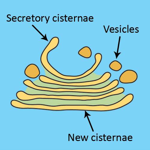
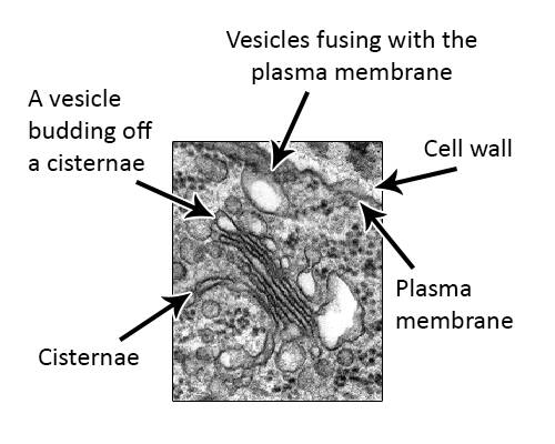
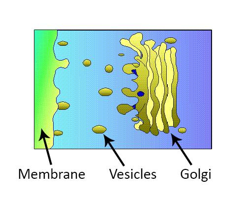
Cell wall materials (mainly polysaccharides) collect in the cisternae and eventually vesicles containing the materials bud off.
Vesicles migrate to the plasma membrane where they fuse with the membrane and empty their contents to the cell wall.
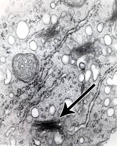
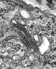
Look carefully at this electron micrograph.
Can you recognize the parts of the golgi body?
Do you see the vesicle depositing material into the cell wall?
See next section