Meiosis is a type of cell division - like mitosis, but it results in four haploid cells with one-half the number of chromosomes as the original diploid cell.
Meiosis occurs in plants only during sexual reproduction in specialized cells to produce a haploid egg cell.
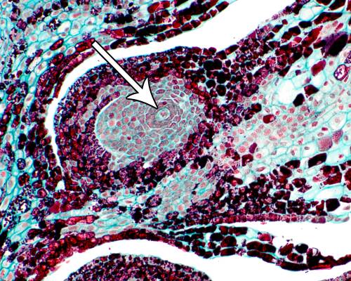
Female egg cell in pine.
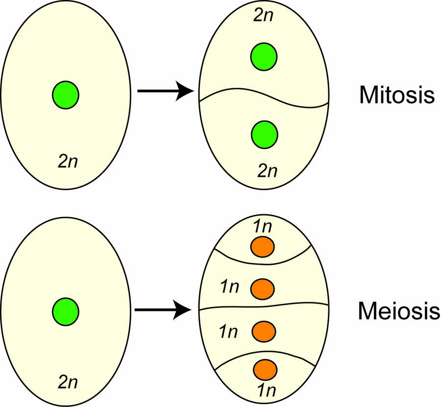
Meiosis differs from mitosis in several important ways.
- The result of a mitotic division is two diploid cells, while meiosis results in four haploid gametes.
- Mitosis requires one cell division, while meiosis requires two divisions.
Meiosis 1
Prophase I
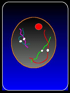
Early in prophase I the chromosomes become visible as thin threads within the nucleus.
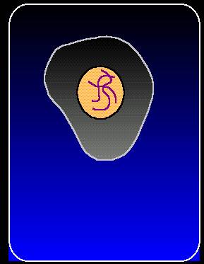
Early in prophase I, the chromosomes become visible as thin threads within the nucleus.
Just as in mitosis, the chromosomes have doubled during interphase and the chromosomes appear as two chromatids attached at the centromere.
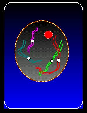
The two chromatids now appear as single condensed threads attached at the centromere.
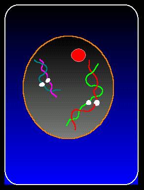
Homologous pairs of chromosomes become associated and are lined up at their centromeres. Each pair is called a bivalent.
An important aspect of prophase I is that the bivalents become tightly intertwined and pieces of one chromatid can cross-over to the other chromosome.
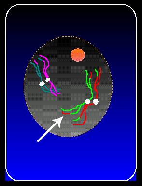
In the last stages of prophase I, you can again see the two chromatids attached to a common centromere.
However, the chromatids are now different because crossing-over has moved genetic material from one homologous chromosome to the other.
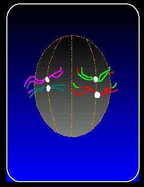
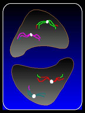
Metaphase I
During metaphase I, the paired chromosomes move to the middle of the cell in preparation for division.
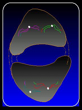
Anaphase I
In anaphase I, the chromosomes separate and move to opposite ends of the cell.

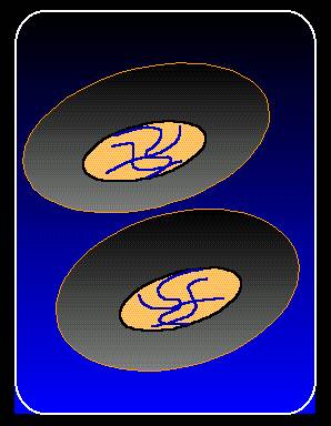
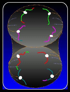
Telophase I
In telophase I, the cell divides and the chromosomes again appear thread-like.
Later in telophase I, cells divide, but no cell plate is made as occurs in mitosis.
Meiosis 2
Prophase II
Prophase II starts the second division stage of meiosis. The chromosomes become more distinct again.
Each chromosome has two chromatids, but notice how each chromatid is no longer identical because of crossing-over.
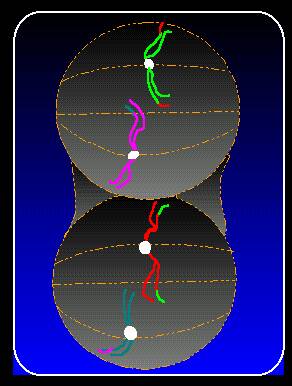
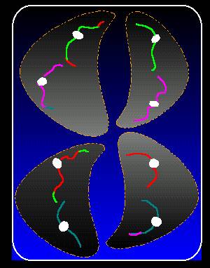
Metaphase II
In metaphase II, the chromosomes line up in the center of the cell.
Anaphase II
In anaphase II, the chromatids pull away from each other. Each has its own centromere.
The separated chromosomes move to opposite ends of the cell.
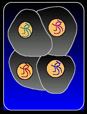
Telophase II
In telophase II, the cells divide, new cell walls are formed and there are four haploid cells. These four cells are called a tetrad.
In telophase II, the cells divide, new cell walls are formed and there are four haploid cells. These four cells are called a tetrad.
Meiosis takes place in the reproductive cells in the flower.
The result of meiosis in the female megagametophyte is an ovule typically with 8 haploid nuclei.
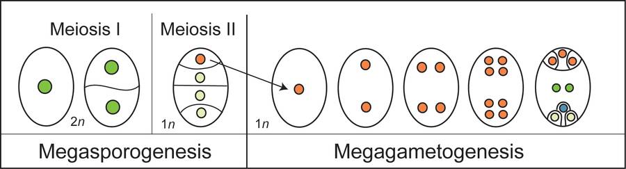
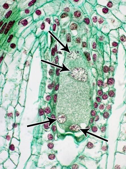
Haploid nuclei in a lily embryo sac.
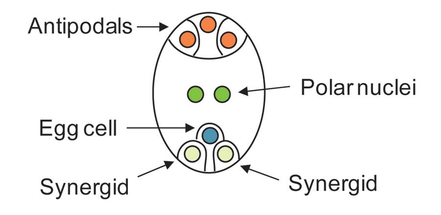
Typical arrangement of nuclei in an embryo sac. A male nucleus will fuse with the polar nuclei to form the endosperm and another with the egg cell to form the embryo.
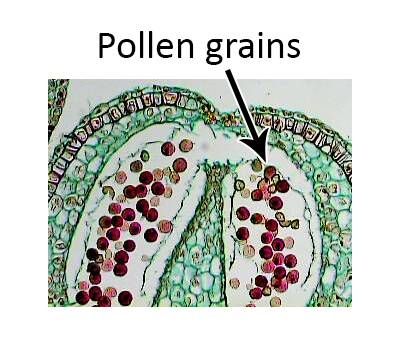
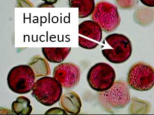
Meiosis in the male part of the flower leads to the production of sperm cells located in the pollen grains. After flower pollination the haploid sperm cell fuses with the female egg cell leading to a fertilized diploid cell that grows into the embryo located in the seed.