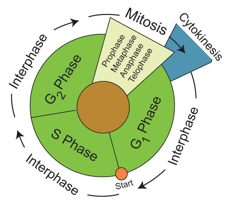
The vegetative cell cycle is divided into two phases.
Interphase and Mitosis
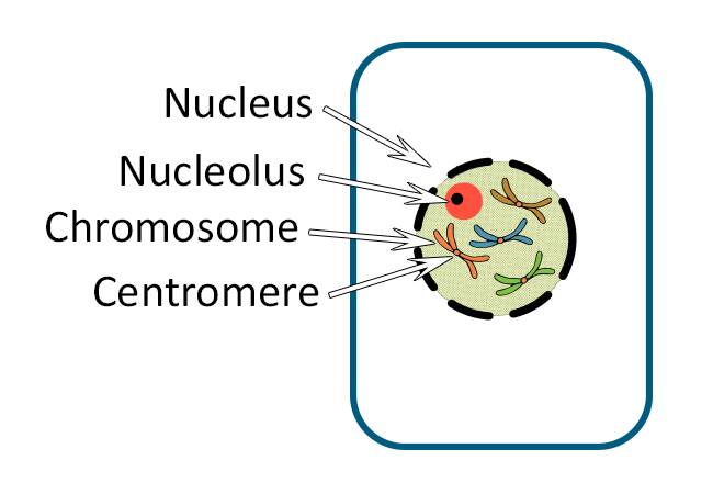
However, many cells in the plant are not actively dividing and are arrested in interphase.
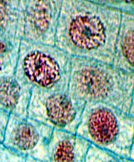
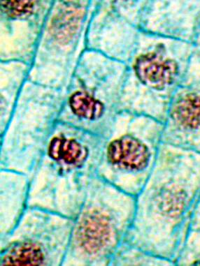
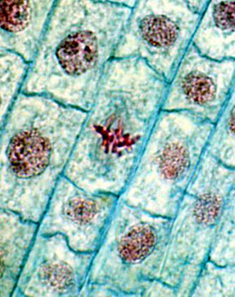
Meristematic cells (like those found in the shoot tip or root tip) are actively cycling from interphase to mitosis as cells divide.
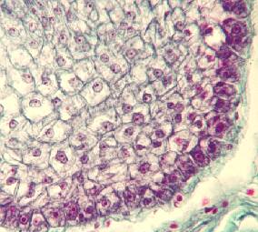
Root Meristem
Interphase is represented as the:
- G1 Phase
- S Phase
- G2 Phase
G1 is the usual resting state for most cells. "G" stands for gap.
Once the cell passes the start point it is committed to cell division.

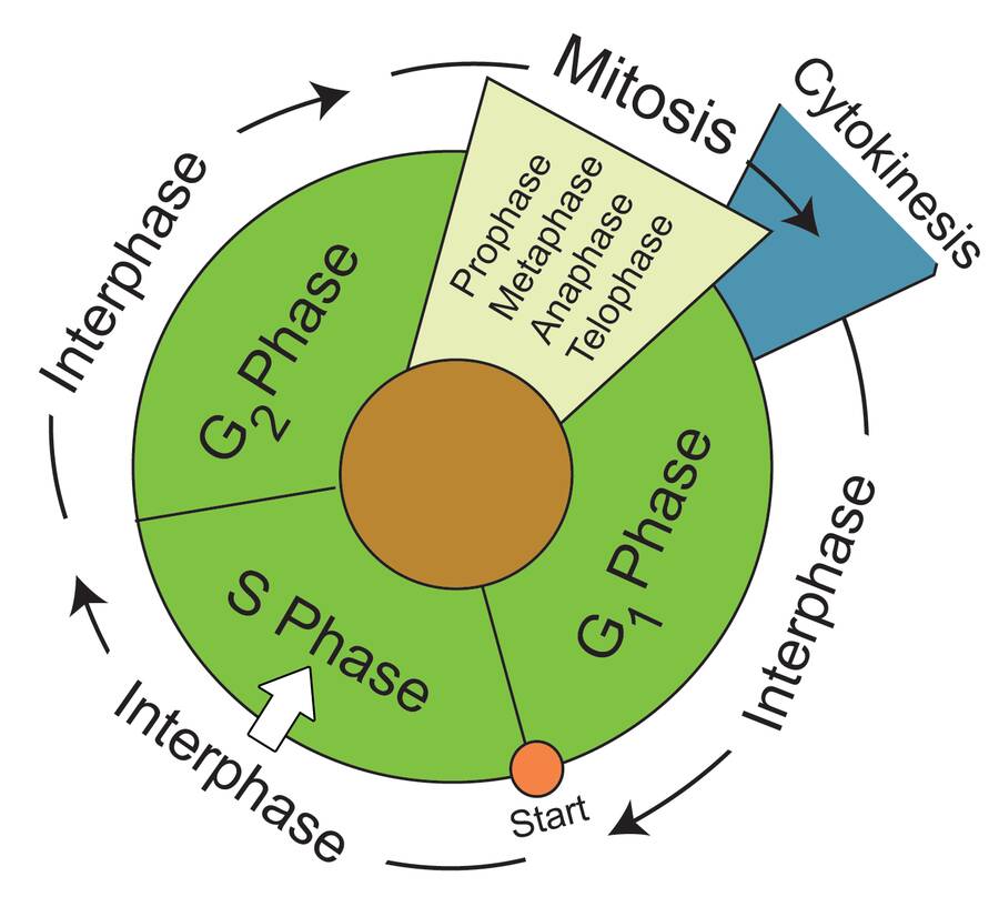
During the S phase both DNA and DNA related proteins are made. This is the phase where a duplicate copy of the cell's DNA is made.
"S" represents synthesis.
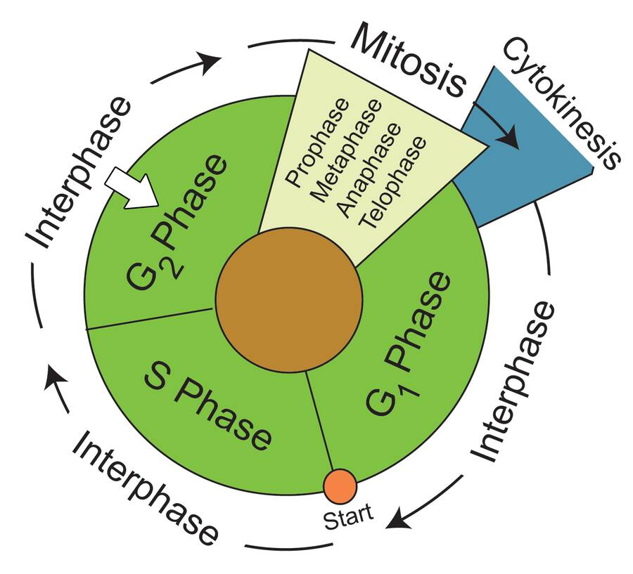
During the G2 phase the cell prepares to divide.
Mitosis is the phase where the cell actually divides. There are four phases:
- Prophase
- Metaphase
- Anaphase
- Telophase
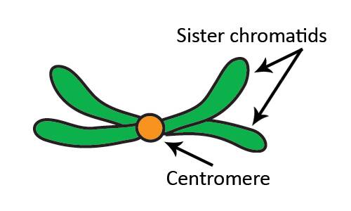
During interphase the chromosomes were duplicated.
These chromosome parts both remain in close association.
At this time, the chromosome consists of two chromatids held together at the centromere.
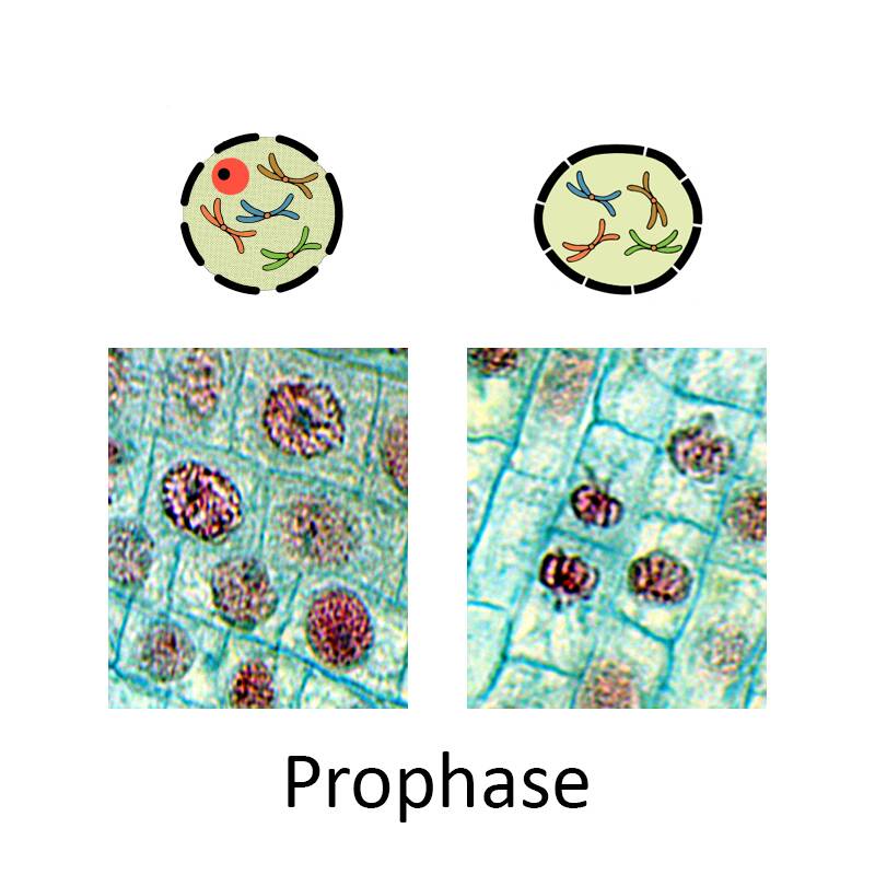
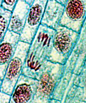
Prophase
During prophase, the chromosomes condense to become well defined.
During late prophase, the nuclear envelope's outer membrane dissolves.
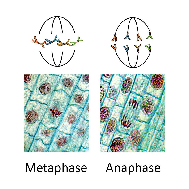
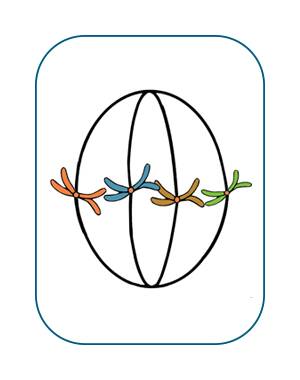
Metaphase
During metaphase, the spindle appears and the chromosomes migrate to the center of the former nucleus.
Anaphase
Spindle fibers become attached to specialized proteins on the centromeres of each sister chromatid.
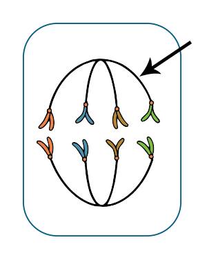
In anaphase, the spindles contract to pull the sister chromatids to opposite sides of the cell. Each is now considered a daughter chromosomes.
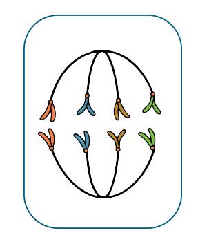
During anaphase, the sister chromatids separate into daughter chromosomes.
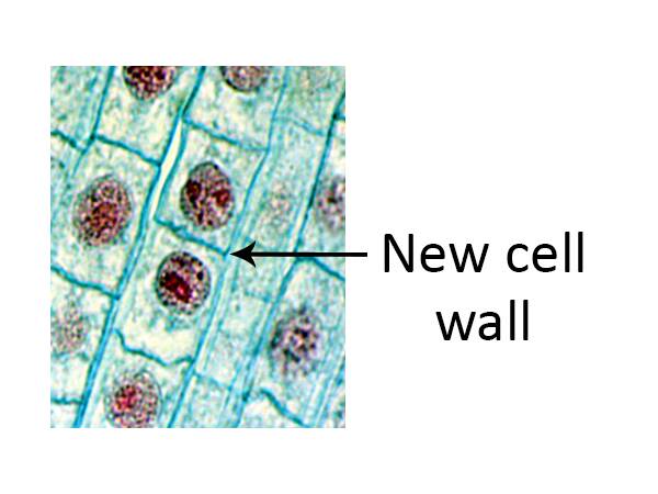
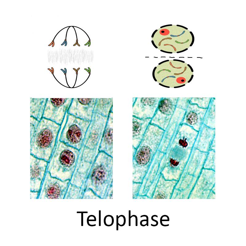
Telophase
During telophase, the chromosomes migrate to their separate cells and the cell plate begins to form.

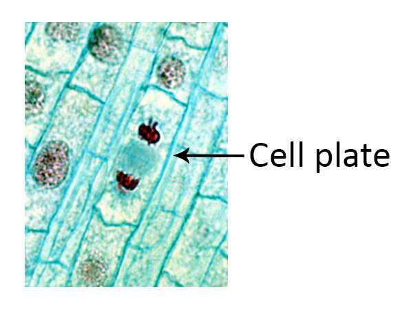
Finally, the cells separate into duplicate daughter cells.